Types Of Veins
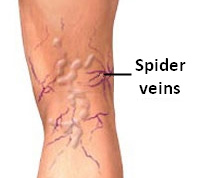 Spider veins are also known as telangiectasias and occur at the earliest stages of venous insufficiency. They are formed by small skin veins that become dilated and are visible through the skin’s surface. They can be blue, purple or red and are frequently extensive, forming various patterns in a linear, starburst, or tree-like distribution. In the legs, they may be associated with varicose veins and venous insufficiency. They can be present in all areas of the body, including the face and the back.
Reticular veins are considered a more advanced stage of a venous disorder. They are formed by larger veins that are located deeper in the skin’s surface with venous reflux. They are usually blue and purple in color, tortuous frequently feeding the areas of spider veins. In contrast to varicose veins, both spider and reticular veins do not protrude on the skin surface. Reticular veins may form areas of unattractive clusters of veins predominantly located on the inner part of thighs, backs of legs and ankles. They can also be associated with symptoms of pain and discomfort of the legs.
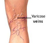 Varicose veins are abnormal veins that become large and tortuous. They appear as rope-like structures protruding out from the skin and can occur throughout the legs and ankles.
Varicose veins occur due to “venous reflux.” Healthy veins have tiny leaflets inside of them called valves. They allow blood to flow in one direction from the ankles towards the heart. When valves do not work, blood backs up in the legs and causes increased pressure in the veins. This causes the veins to enlarge and causes symptoms such as pain, heaviness, cramping, itching and burning of the skin. In advanced cases, this can lead to skin pigmentation, ulceration and bleeding.